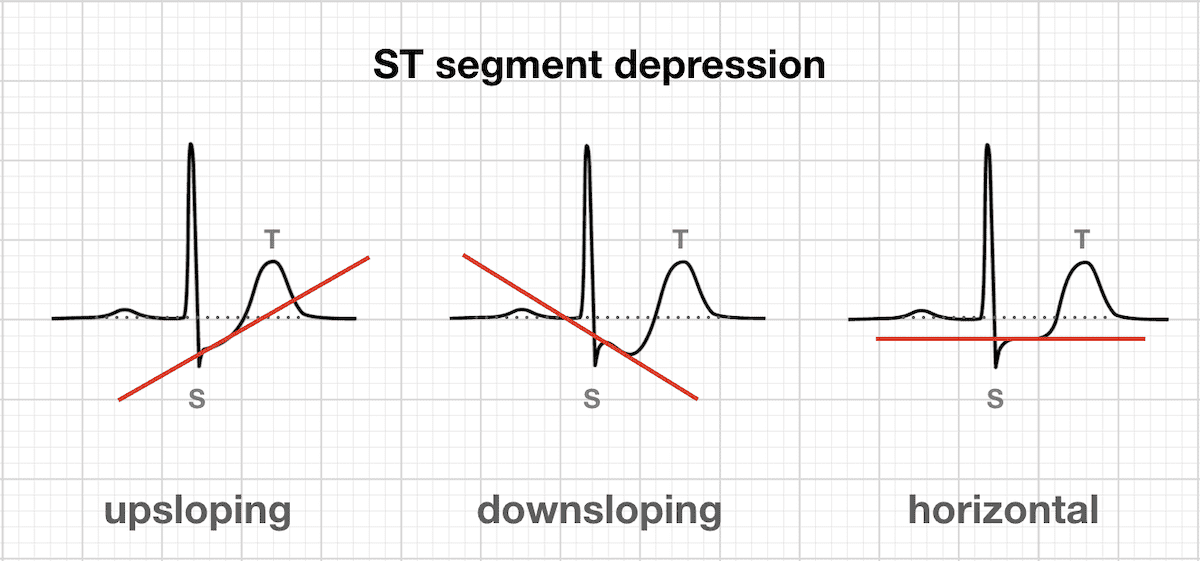
We enrolled 33 patients group 1 with rapid upsloping ST depression 1 mm extending 008 seconds beyond J point 32 patients group 2 with slow upsloping depression 15 mm extending 008 seconds beyond J point and 35 patients group 3 with horizontal or downsloping depression 1 mm at 008 seconds beyond J point. Possible causes include Digitalis Toxicity.
 Stemi St Elevation Myocardial Infarction Diagnosis Criteria Ecg Management Ecg Echo
However these terms do not indicate defined abnormalities but are only descriptive.
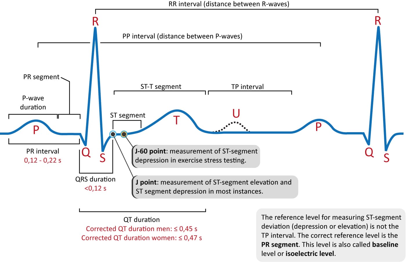
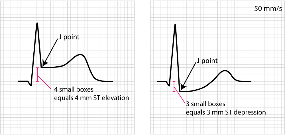
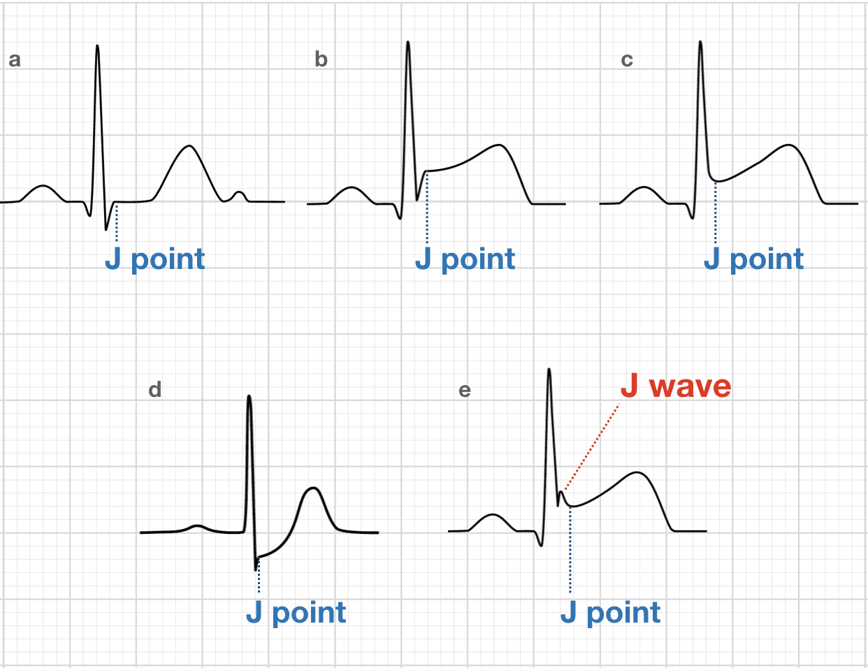
J point depression causes. 10232017 J point depression of 2 to 3 mm in leads V4 to V6 with rapid upsloping ST segments depressed approximately 1 mm 80 milliseconds after the J point. The letter J on the ECG defines 2 totally different and unrelated eventsThe J point is a point in time marking the end of the QRS and the onset of the ST segment present on all ECGs. 242021 Elevation or depression of the J point is seen with the various causes of ST segment abnormality.
With postpartum depression mothers develop symptoms of depression. ESTNot all types of ST segment are pathologicalThe ST segment should depress atleast 1 mm below the isoelectric segment and it should be depressed for 80msec from the J point. Summed stress score SSS.
Upsloping ST depression J point depression -. Talk to our Chatbot to narrow down your search. The terms J point elevation and J point depression often cause confusion among trainees who mistakenly think that these terms denote a specific condition.
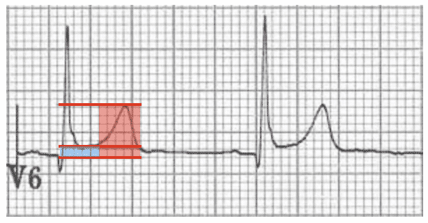
Check the full list of possible causes and conditions now. The right panel of Figure 4 shows an upsloping ST depression with depressed J-60 point and J-80 point. Osborn wave J wave.
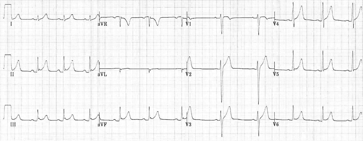
The junction J point is where the QRS complex and ST segment meet. The J wave is a much less common long slow deflection of uncertain origin originally. Malignant variant associated with arrhythmias and sudden cardiac death typically has inferolateral J wave notching or slurring at the end of the QRS.
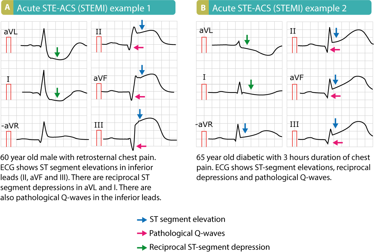
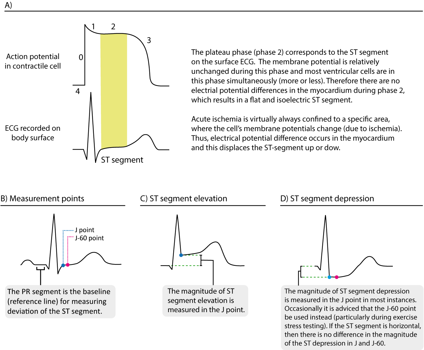
J waves in multiple leads esp. Anemia metabolic abnormalities MVP normal variant. The most important cause of ST segment abnormality elevation or depression is myocardial ischaemia or infarction.
E with J wave Osborn wave Note. Causes They are usually observed in people suffering from hypothermia with a temperature of less than 32 C 90 F 5 though they may also occur in people with high blood levels of calcium hypercalcemia brain injury vasospastic angina acute pericarditis or ventricular fibrillation and could also be a normal variant. 942009 ST segment depression is the classical response to stress during excercise stress testing.
The ST-segment slope in leads V4 and V5 is 30 mVsec. B c J point elevation. It must satisfy two criteria.
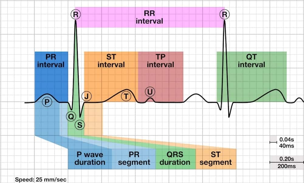
5282020 J point in a normal. The ST segment is the flat isoelectric section of the ECG between the end of the S wave the J point and the beginning of the T wave. The ST Segment represents the interval between ventricular depolarization and repolarization.
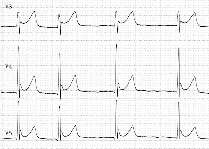
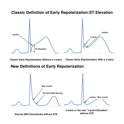
Such ST depressions are also common during exercise and situations with tachycardia. Possible causes include Digitalis Toxicity. For example isolated J point elevation may occur as a normal variant with the early repolarization pattern see Chapter 10 or as a marker of systemic.
Causes widespread concave STE with notching or slurring at the J-point fishhooks J waves Benign variant typically causes mostly precordial STE. Orthostatic Hypotension Symptom Checker. Talk to our Chatbot to narrow down your search.
Inferior or with higher amplitudes 02 mV are. It may be elevated as a result of injury currents during acute myocardial ischemia and pericarditis as well as in various other patterns of both normal and abnormal ECGs. Elevation of the J point occurs with benign early repolarisation.
These waves occur due to hypothermia hypercalcemia early repolarization and Brugada syndrome. J-Point Depression Symptom Checker. The quantum of ST depression should.
8112020 This affects the plateau phase of the ventricular transmembrane action potential and hence the ST segment. ST depression can also be seen in infarction typically in non Q-wave infarction often called subendocardial infarction. In cases in which myocardial injury has occurred if ST elevation is present leads electrically opposite tend to show reciprocal ST depression.
J point depression at the beginning of the QRS complex is not significant if the location of measurement two boxes past the QRS finds the ST segment has risen back to the baseline. D J point depression. ST usually back to baseline 2 mm after the end of QRS.
There are four principial causes of J waves namely hypothermia Brugada syndrome early repolarization and hypercalcemia. 122020 Any changes in hormone states including menopause childbirth thyroid problems or other disorders could cause depression. In summary J point depression is not caused by ischemia.
Check the full list of possible causes and conditions now.
 Benign Early Repolarisation Litfl Ecg Library Diagnosis
 Myocardial Ischaemia Litfl Ecg Library Diagnosis
 Ecg Interpretation Characteristics Of The Normal Ecg P Wave Qrs Complex St Segment T Wave Ecg Echo
J Point Ecg Interval Litfl Ecg Library Basics
 Benign Early Repolarisation Litfl Ecg Library Diagnosis

 The St Segment Physiology Normal Appearance St Depression St Elevation Ecg Echo
 Stemi St Elevation Myocardial Infarction Diagnosis Criteria Ecg Management Ecg Echo
 Benign Early Repolarisation Litfl Ecg Library Diagnosis
 Myocardial Ischaemia Litfl Ecg Library Diagnosis
 The St Segment Physiology Normal Appearance St Depression St Elevation Ecg Echo
 Osborn Wave J Wave Litfl Ecg Library Basics
 J Point Ecg Interval Litfl Ecg Library Basics
 Benign Early Repolarisation Litfl Ecg Library Diagnosis
 Osborn Wave J Wave Litfl Ecg Library Basics
 J Point Ecg Interval Litfl Ecg Library Basics
 Early Repolarization Ecgpedia
 Benign Early Repolarisation Litfl Ecg Library Diagnosis
 Benign Early Repolarisation Litfl Ecg Library Diagnosis


