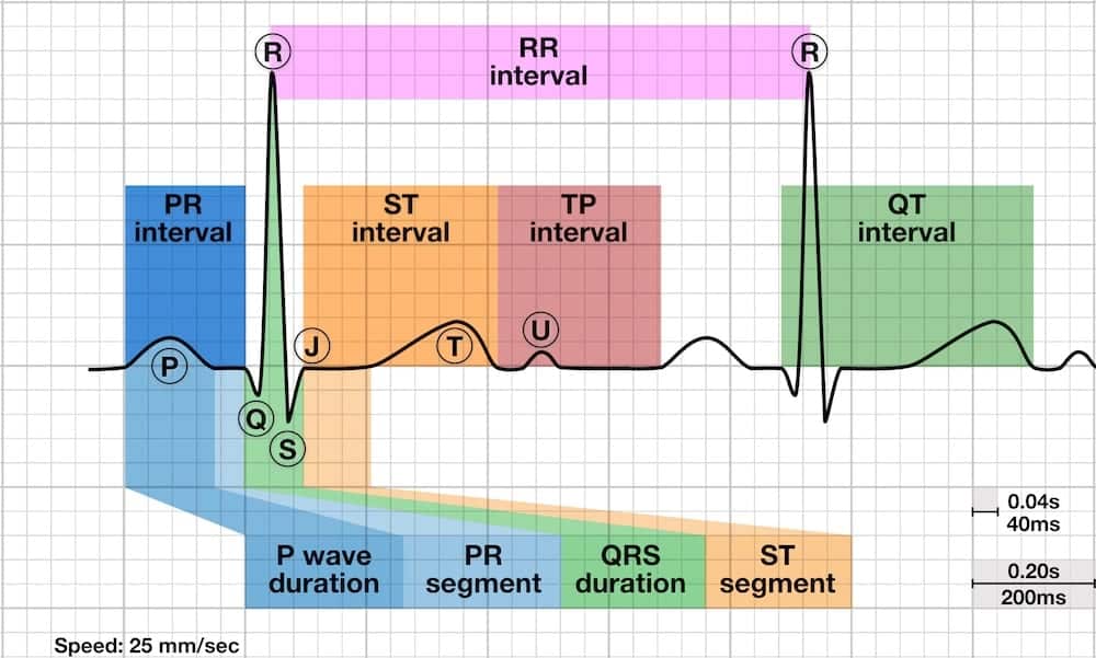
The direction of the wave above or below the isoelectric line. Acute Pleuritic Chest Pain.
 Diagnosis Of Acute Pericarditis
Reciprocal ST depression and PR elevation in lead aVR.

P wave depression pericarditis. Only STEMI causes convex-up or horizontal ST elevation. PTa depression depression between the end of the P-wave and the beginning of the QRS- complex stage II. PR depression only reliably seen in viral pericarditis and often only an early transient phenomenon lasting hours.
PTa depression depression between the end of the P wave and the beginning of the QRS complex Stage II. 142012 Pericarditis does not produce abnormal Q waves as seen with myocardial infarcts. Absence of reciprocal ST depression elsewhere.
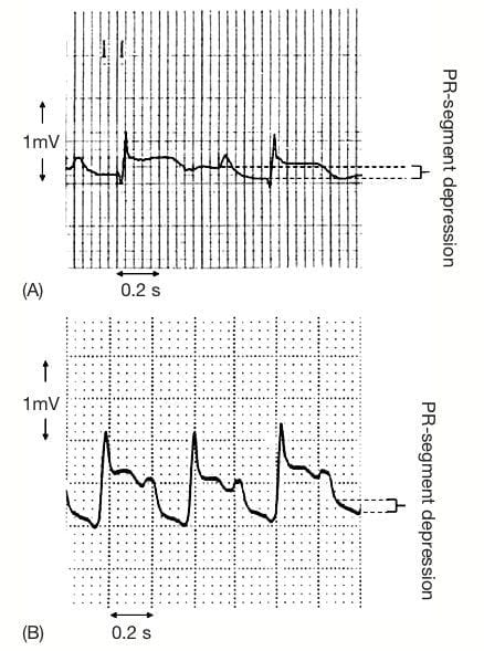
At stage 1 ECG findings are very similar to those of the early repolarization. PR depression or PR elevation in lead aVR can also be seen in an atrial infarction. Depression of the PR segment is very specific of acute pericarditis and is attributed to subepicardial atrial injury and occurs in all leads except aVR and V1.
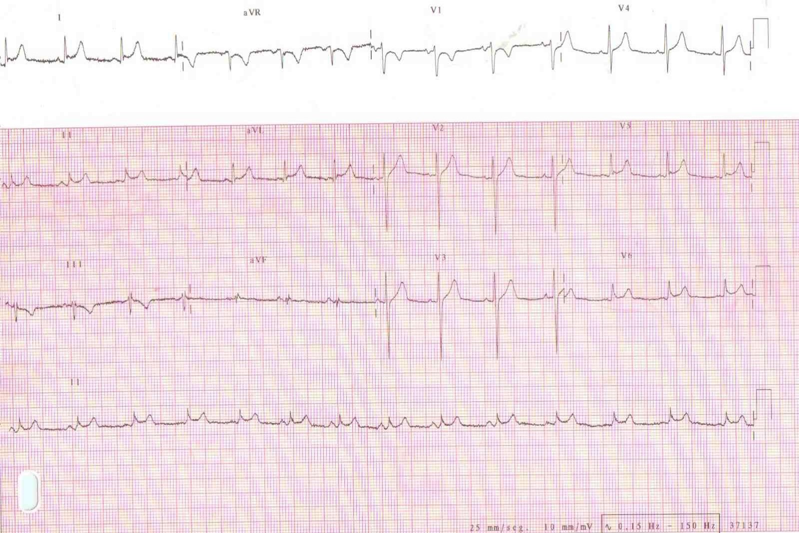
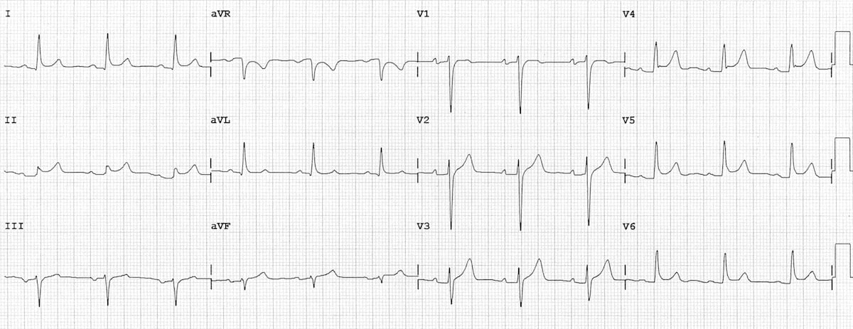
In pericarditis four stages can be distinguished on the ECG. Such depressions occur in most leads except from lead V1 which often shows PR segment elevation. And Gettes LS 2009.
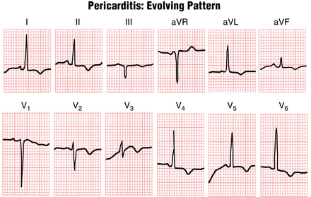
ST elevation in all leads. Widespread concave saddle-shaped ST elevation. Pericarditis causes only superficial inflammation but not frank myocardial necrosis.
The characteristic changes of acute pericarditis are. Abnormal Q waves are absent in pericarditis but they are present in myocardial infarction. Shown below is an EKG image of pericarditis at several different stages.
Diffuse ST segment elevation disappears. STEMI can have concave-up ST elevation like pericarditis. The pattern of ST-segment elevation is important in the diagnosis of acute pericarditis.
PR segment changes are relative to the baseline formed by the T-P segment. Diffuse concave upward ST segment elevation in most leads PR depression in most leads may be subtle and sometimes notching at the end of the QRS complex. 1122015 P Waves in the Normal ECG P waves are described according to.
PR segment depression or elevation PR segment deviates opposite to the polarity of P wave. Rautaharju PM Surawicz B. AHAACCFHRS recommendations for the standardization and interpretation of the electrocardiogram.
The ST segment T and U waves and the QT interval a scientific statement from the American Heart Association Electrocardiography and. They are called upright or positive P waves when the deflections are above the isoelectric line or are called downward or negative or inverted P waves when the deflections are below the isoelectric line. Elevation or depression of the PTa segment the part between the p wave and the beginning of the QRS complex can result from atrial infarction or pericarditis.
Stage I acute phase. If the P wave is inverted it is most likely an ectopic atrial rhythm not originating from the sinus node. Upwardly concave ST segment elevation in all leads except aVR.
1122011 The Abnormal P wave. The abnormal Q wave in myocardial infarction is due to death of heart muscle which leads due to loss of positive depolarization voltages. 812020 Pericarditis is classically associated with ECG changes that evolve through four stages.
Pseudonormalization transition Stage III. 1112002 Pericarditis or inflammation of the pericardium is most often caused by viral infection. Reciprocal ST depression and PR elevation in aVR and V1.
Although PQ segment depression is known to be one of the typical ECG changes associated with acute pericarditis 5 6 the incidence and clinical significance of PQ segment depression in patients with acute Q wave inferior myocardial infarction have not been established. Disease the P wave and QRS complexes are normal. The PR segment elevation and depression The PR segment is not affected in STEMI whereas acute pericarditis often causes PR segment depression.
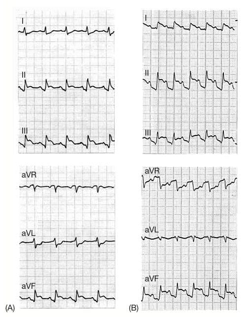
The ECG changes in pericarditis are generally observed at 4 stages. 3202021 Diffuse PR segment depression affecting virtually all leads except in leads V1 and aVR is a quasispecific finding in acute pericarditis but it is absent in STEMI. Sinus tachycardia is also common in acute pericarditis due to pain andor pericardial effusion.
Stage 1 widespread STE and PR depression with reciprocal changes in aVR occurs during the first two weeks Stage 2 normalisation of ST changes. ST elevation in all leads. If the p-wave is enlarged the atria are enlarged.
Generalised T wave flattening 1 to 3 weeks Stage 3 flattened T waves become inverted 3 to several weeks. Pseudonormalisation transition stage III. Widespread concave ST elevation and PR depression throughout most of the limb leads I II III aVL aVF and precordial leads V2-6.
-segment depression Upward concave ST-segment elevation No T-wave. In our study 14 of 171 patients 8 with Q wave inferior wall acute myocardial infarction had PQ segment depression. These leads may exhibit PR-segment elevation 14.
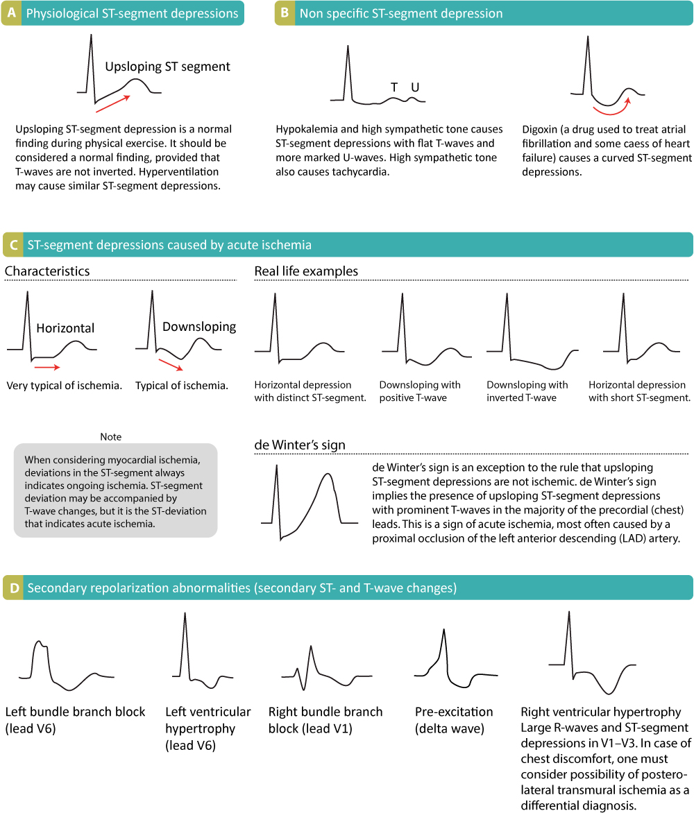 St Segment Depression In Myocardial Ischemia And Differential Diagnoses Ecg Echo
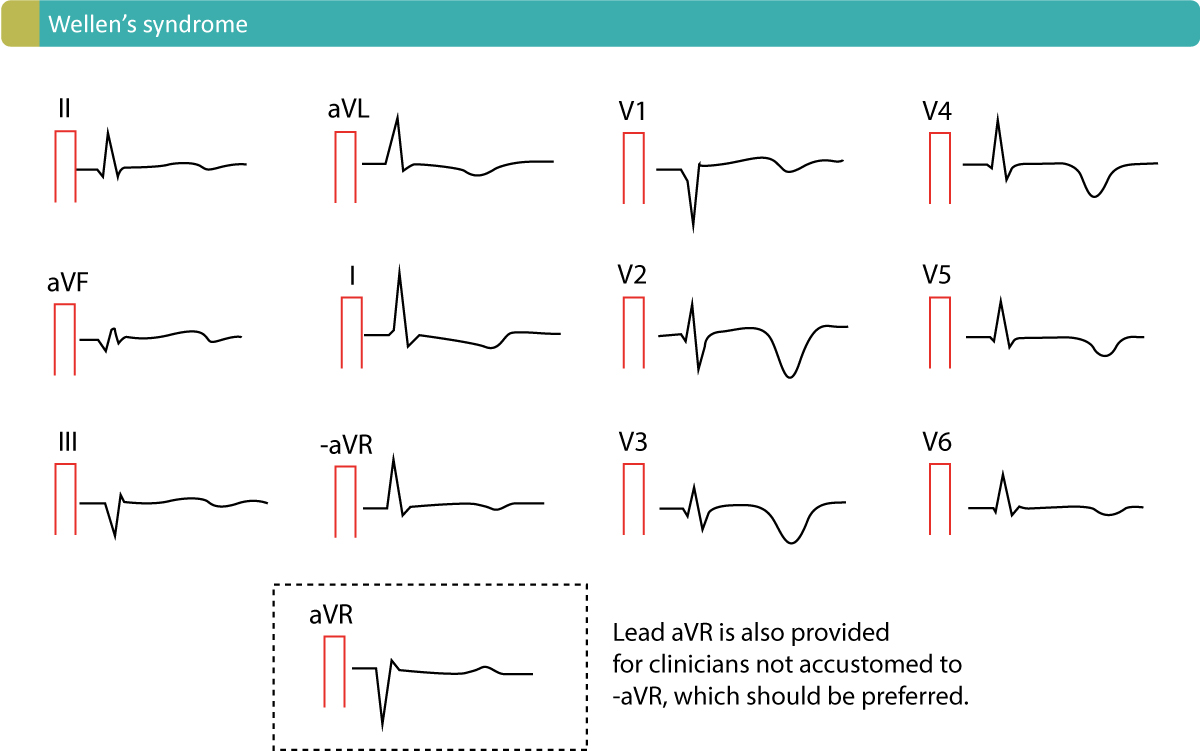 T Waves In Ischemia Hyperacute Inverted Negative Wellen S Sign De Winter S Sign Ecg Echo
 Dr Smith S Ecg Blog Pericarditis Strikes Again
 Ecg Pericarditis Ecg Abnormalities Suggestive Of Pericarditis In A 40 Download Scientific Diagram
 A Schematic Illustration Of The Possible Electrocardiographic Ecg Download Scientific Diagram
 Pr Segment Litfl Ecg Library Basics
 Pr Segment Litfl Ecg Library Basics
 Ecg Pericarditis Ecg Abnormalities Suggestive Of Pericarditis In A 40 Download Scientific Diagram
 A Ecg Showing Concave St Elevation And Pr Segment Depression In All Download Scientific Diagram
 Left Atrial Enlargement P Mitrale Right Atrial Enlargement P Pulmonale On Ecg Ecg Echo
 Dr Smith S Ecg Blog You Diagnose Pericarditis At Your Peril At The Patient S Peril
 Pr Segment Litfl Ecg Library Basics
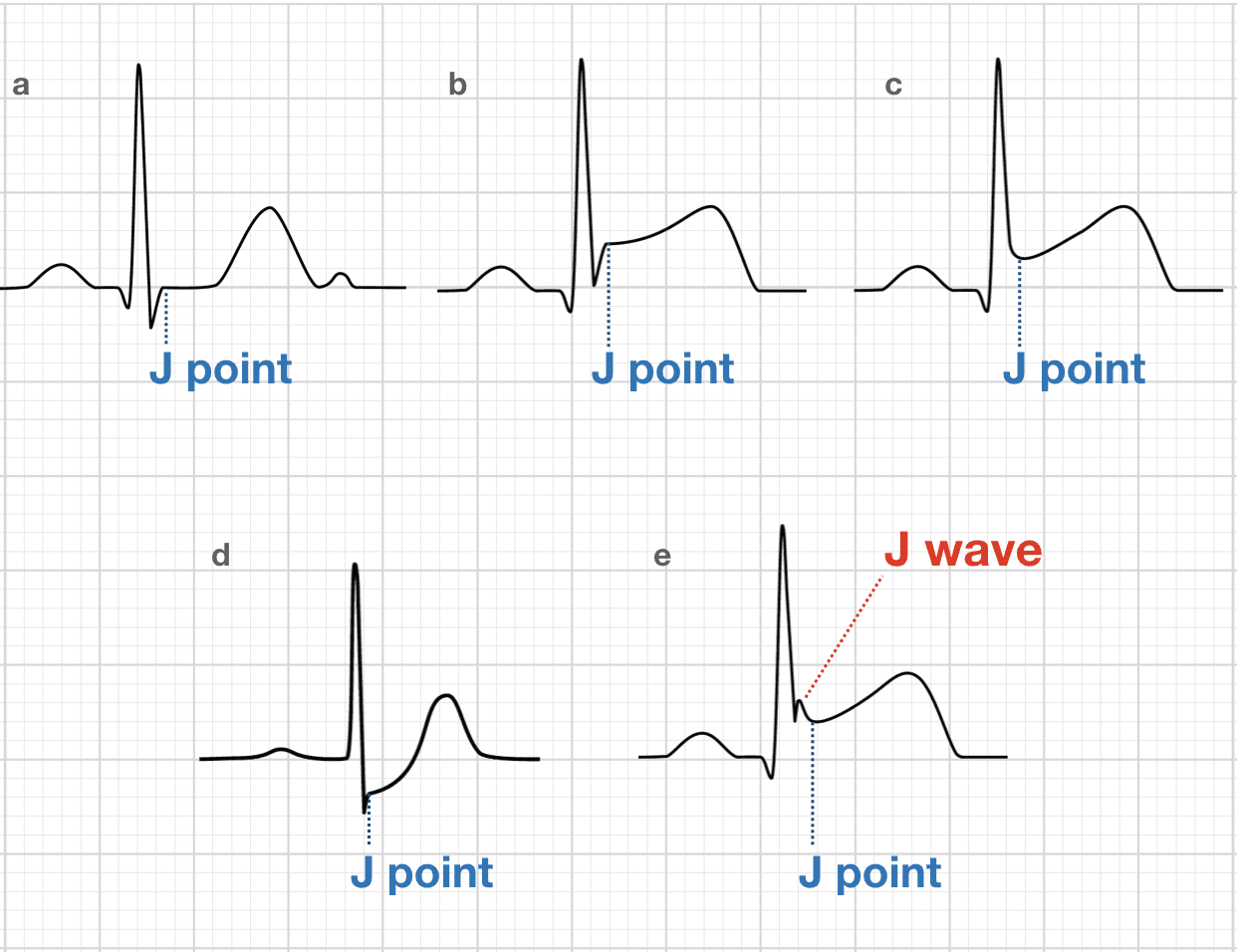 J Point Ecg Interval Litfl Ecg Library Basics
 Acute Pericarditis Diagnosis And Management American Family Physician
 Dr Smith S Ecg Blog Pericarditis Strikes Again
Ecg Waveforms Ecg Techniques And Recognition
 Dr Smith S Ecg Blog Pericarditis Strikes Again
 Pr Segment Litfl Ecg Library Basics
 Pericardial Myocardial And Pulmonary Syndromes Thoracic Key


