The electrical findings from your exercise treadmill test ie. ST 05 mm is a better threshold than 10 mm to define ECG evidence for regadenoson-induced myocardial ischemia.
 Dr Smith S Ecg Blog Deep And Widespread St Depression Signifies High Risk Coronary Lesion
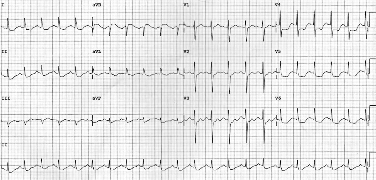
172009 Notice the very subtle ST depression in III and aVF.

St depression 0.5 mm. Hope this information was useful to you. It could be persistent or transient and it is a sign of disturbances during. 1212004 Patients were excluded if they presented with one or more of the following features.
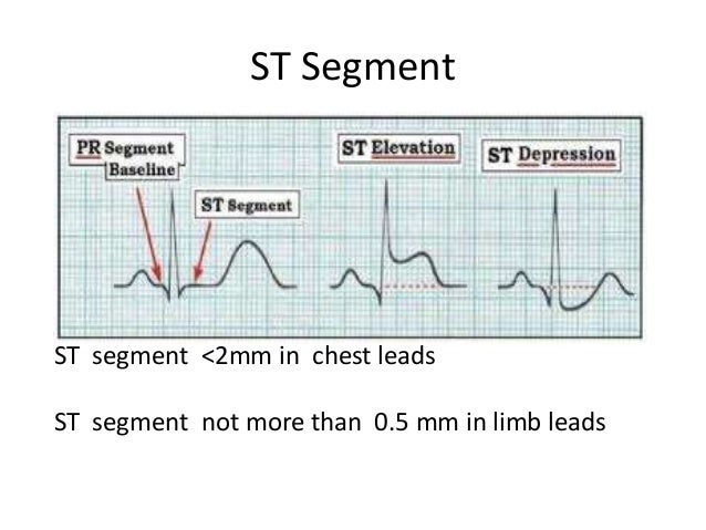
ST depression 1 mm is more specific and conveys a worse. At rest there is a normal ST segment upper panel but during an increase in heart rate lower panel 1 mm horizontal ST depression ensues. 3202021 ST-Segment Depression and Myocardial Ischemia.
Current guideline criteria for ischemic ST segment depression. Two patients with ST depression both in the strict criteria group had dyspnea during the ST segment depression that was only reported after we asked confirmation of the absence of complaints in the diary. Normal range at rest is between 60-100 beats per minute bpm.
8222018 ST segment depression less than 05 mm is accepted in all leads. 1 mm 01 mV. And clinical evidence of non.
ST depression of 15mm is not significant considering your age But still for complete Evaluation I recommend a TMT test for you. ECG abnormalities at rest that could have interfered with ST segment analysis including atrial fibrillation baseline ST segment depression. With 15mm you are a little below that but with this depression you are still suspect for ischemia.
However although ST depression often comes from ischemia there may be other causes for it. 212020 Isolated ST-segment depression of at least 05 mm in leads V1V3 is the primary observational finding of an AMI within the inferior and basal portion of. Thus it seems that perhaps there may be 05-10 mm STE in aVR without any ST depression of at least 05 mm.
132010 Furthermore the vast majority of patients with STE in aVR had between 05 and 10 mm. An ST depression gives better indication. Example of left ventricular hypertrophy with typ-ical secondary ST-T abnormalities in leads I II aVL V 4 V 5 and V 6.
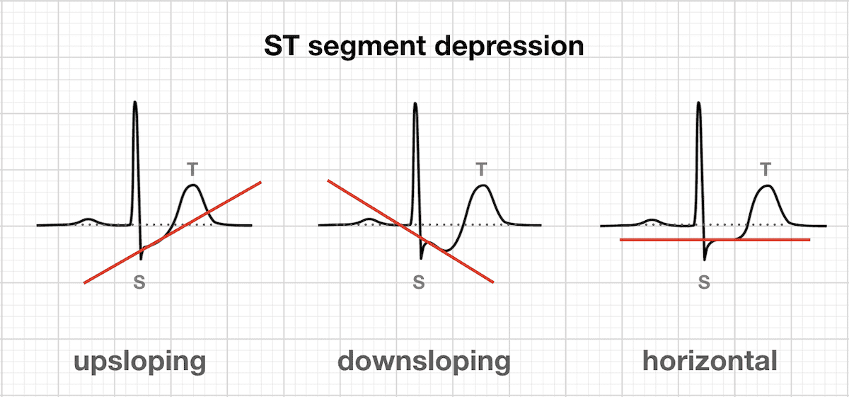
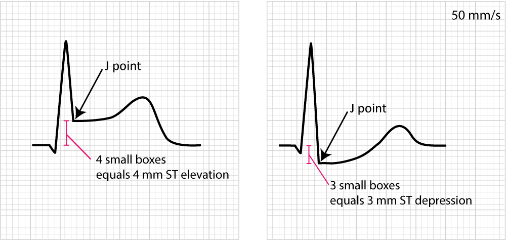
ST depression can be either upsloping downsloping or horizontal see diagram below. Some expert consensus documents also note that any ST segment depression in V2V3 should be considered abnormal because healthy individuals rarely display depressions in those leads. New horizontal or downsloping ST segment depressions 05 mm in at least two anatomically contiguous leads.
5 mm 05 mV or between 2 dark horizontal lines 10 mm 10 mV. 6 months acute MI. The criterion of 2 mm of additional exercise-induced ST-segment depression or downsloping depression of 1 mm or more in recovery was a particularly useful marker for the diagnosis of any coronary disease likelihood ratio 34 sensitivity 67 and specificity 80.
Lead III also shows ST depression. Therefore your doctor ordered the. Any st-segment depression or elevation 05 mm may be abnormal Figure 2.
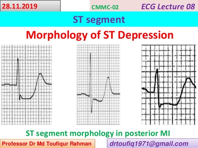
812020 Morphology of ST Depression. ST segment depression 05 mm or more is considered pathological. Kindly get it done and revert back.
That is the reason why your doctors need to study your heart in more detail with a heart cath coronary angiogram. ST segment depression can be Ischemia as in exercise EKGs or subendocardial injury current. 05 mm left bundle branch block and pacemaker rhythm.
The rule is that 2 mm or more significantly indicates reversible ischemia. 05 mm ST depression and T inversion during recovery are non-specific so they do not necessary mean that you have an artery of the heart occluded. Horizontal or downsloping ST depression 05 mm at the J-point in 2 contiguous leads indicates myocardial ischaemia according to the 2007 Task Force Criteria.
The transition from ST segment to T-wave is more abrupt in ischemia the transition is. HI Greetings from DrDivakaraP Thanks for posting your query. AVF circled has 05 mm of ST depression in the context of a 1 mm QRS ST depression should be considered with proportionality in mind.
Regadenoson-induced ischemic ST is more common in women and it provides a modest independent prognostic value beyond MPI and clinical parameters. However any large amount greater than or equal to 10 mm will have some ST depression. These should be interpreted as reciprocal to aVL the opposite lead.
The QRS complex is upright in these leads while the ST. Acute ST-segment depression is as elevation a sign of myocardial injuryIt generally correlates with incomplete coronary artery occlusion see NSTE-ACSAs with elevation ST-segment depression must be present in at least two adjacent leads.
 Electrocardiogram Showing St Depression In The Inferolateral Leads And Download Scientific Diagram
 Stop Now What You Doing Tomorrow St Depression
 Dr Smith S Ecg Blog Deep And Widespread St Depression Signifies High Risk Coronary Lesion
 Stemi St Elevation Myocardial Infarction Diagnosis Criteria Ecg Management Ecg Echo
 Ecg St Segment
 Dr Smith S Ecg Blog Deep And Widespread St Depression Signifies High Risk Coronary Lesion
 Dr Smith S Ecg Blog Widespread St Elevation Activate The Cath Lab
 Myocardial Ischaemia Litfl Ecg Library Diagnosis
 Electrocardiogram Showed Sinus Rhythm With Upsloping St Segment Download Scientific Diagram
 Myocardial Ischaemia Litfl Ecg Library Diagnosis
 Ecg Theory Presentation 1 Ecg Presentations
 Myocardial Ischaemia Litfl Ecg Library Diagnosis
 St Segment Elevations
 Myocardial Ischaemia Litfl Ecg Library Diagnosis
 Myocardial Ischaemia Litfl Ecg Library Diagnosis
 Myocardial Ischaemia Litfl Ecg Library Diagnosis
 Electrocardiogram Showing St Depression In The Inferolateral Leads And Download Scientific Diagram
Anterior Stemi There Is Upsloping St Elevation In The Anterior Chest Download Scientific Diagram
 The St Segment Physiology Normal Appearance St Depression St Elevation Ecg Echo